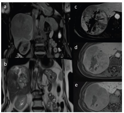
Fig. (19).

Large HCC with degenerative changes in coronal T1-weighted image with fat saturation (a) and in coronal T2-weighted image (b). Dynamic contrast-enhanced sequences in axial T1-weighted images with fat saturation after administration of hepatocyte-specific contrast agent in hepatic arterial phase (c), portal venous phase (d) and hepatobiliary phase (e). Heterogenous enhancement of the lesion is seen with areas of non-enhancing focal necrosis (c) with subsequent washing out of the contrast agent (d). Lesion shows low signal intensity in comparison to adjacent liver parenchyma in hepatobiliary phase (e).
