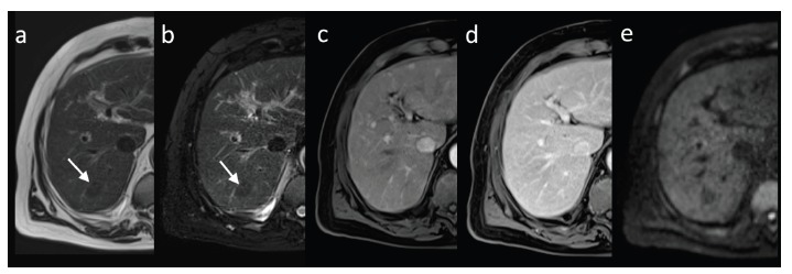
Fig. (2).

MRI, axial image of low grade dysplastic nodule in segment VII (arrow). Nodule is hypointense in T1-weighted (a) and T2-weighted fat-suppressed images (b), shows isointensity to the surrounding liver parenchyma in hepatic arterial phase (c) and in consecutive phase (d); no diffusion restriction in diffusion weighted imaging (b value of 800s/mm2, e) is seen.
