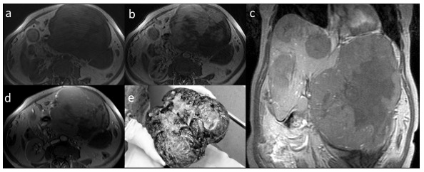
Fig. (4).

MRI, fibrolamellar carcinoma (FLC) in axial T1-weighted images pre-contrast (a), with heterogenic enhancement in hepatic arterial phase (b) and equilibrium phase (d). The mass in coronal plane presents no enhancement in hepatobiliary phase, other foci of FLC are visible (c). Post-operative image of the large mass (e).
