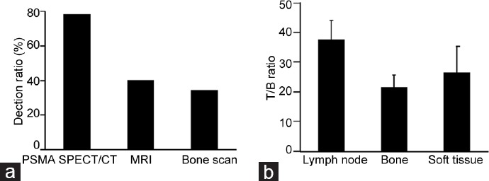
Figure 1.

Comparison of different imaging modalities for metastatic lesions in PCa patients. (a) Prostate-specific membrane antigen (PSMA) single-photon emission computed tomography (SPECT)/computed tomography (CT) imaging provided a higher detection ratio than bone scan or magnetic resonance imaging (MRI). (b) The tumor/background (T/B) ratio was higher for lymph node metastases than soft-tissue or bone metastases.
