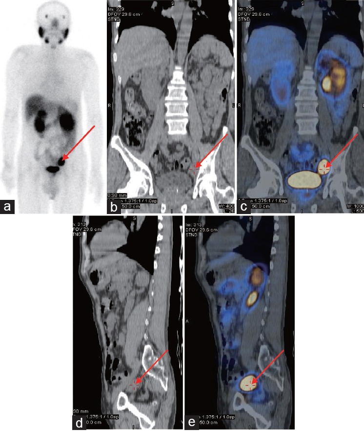
Figure 5.

Example results for a patient who received RP and extended pelvic lymphadenectomy. The patient was 62-year-old male with a biopsy-proven prostate cancer Gleason score of 8 and PSA level of 46 ng ml−1. PSMA-SPECT/CT indicated lymph node metastases near the left iliac blood vessels ([a] Whole-body planar images; [b] coronal plane of CT plain scan; [c] coronal plane of fused PSMA-SPECT/CT images; [d] vertical plane of CT plain scan; [e] vertical plane of fused PSMA-SPECT/CT images.). Following PSMA-SPECT/CT imaging, the patient underwent RP and extended pelvic lymphadenectomy. The PSMA-positive lesions near the left iliac blood vessels were proven to be prostate cancer using histology. After surgery, the patient received ADT and his PSA level decreased to 0.10 ng ml−1. Arrows indicate metastatic lymph node lesions detected using PSMA-SPECT/CT.
