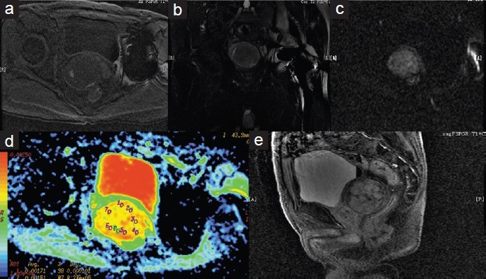
Figure 2.

On cross section T1-weighted image (a), the tumor is mixed hypointense and hyperintense. On coronal T2-weighted (b) shows mixed hperintense signal tumor with hypointense capsule. On cross section, DW image (c) shows pale hyperintense signal. On cross section ADC image (d) shows the ADC is about 1.68 × 10⁻³ mm2 s−1. Contrast-enhanced MRI scan (e) shows prolonged and delayed enhancement pattern with tumor capsule. DW: diffusion-weighted; ADC: apparent diffusion coefficient; MRI: magnetic resonance imaging.
