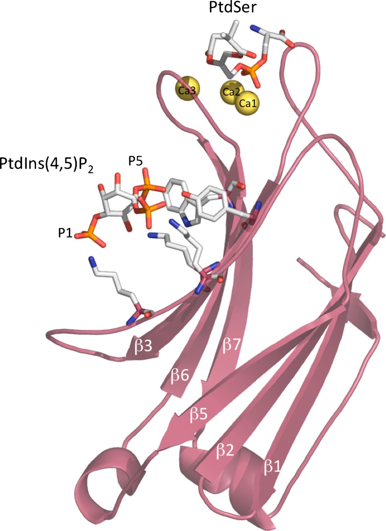
Fig. 3.
Structure of PKCα-C2 domain bound to Ca2+-phosphatidylserine–phosphatidylinositol-4,5-bisphosphate in a quaternary complex. The C2 molecule is shown in magenta. The three calcium ions are shown in yellow spheres, with one of these spheres bridging the protein with PtdSer at the tip of the domain. The PIP2 molecule is bound to the β3–β4 chains (Guerrero-Valero et al. 2009). Protein Data Bank (PDB) accession number 3GPE

