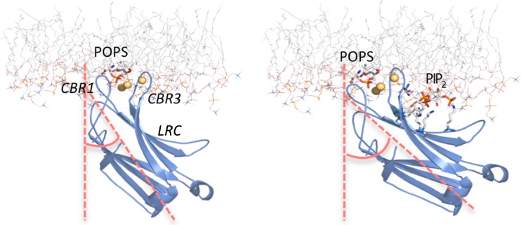
Fig. 4.
C2 domain of PKCα docked onto the surface of membranes without (left) and with (right) PIP2. The C2 domain of PKCα is represented as a ribbon model in blue by using the PDB codes 1DSY(left) (Verdaguer et al. 1999) and 3GPE (right) (Guerrero-Valero et al. 2009). The lipid membrane corresponds to a phosphatidylcholine (POPC) molecular dynamics simulation (POPC128A) (Hoff et al. 2005) and is represented as a stick model with carbon in gray, nitrogen in blue and oxygen in red. The headgroups of the two anionic phospholipids, phosphatidylserine (POPS) and PIP2 led us to dock the domain in the hydrophilic region of the membrane bilayer. It can be observed that in the presence of PIP2, the angle formed by the axes of the protein with the normal to the bilayer increases. Figure prepared with PyMOL (DeLano Scientific LLC, San Francisco, CA)

