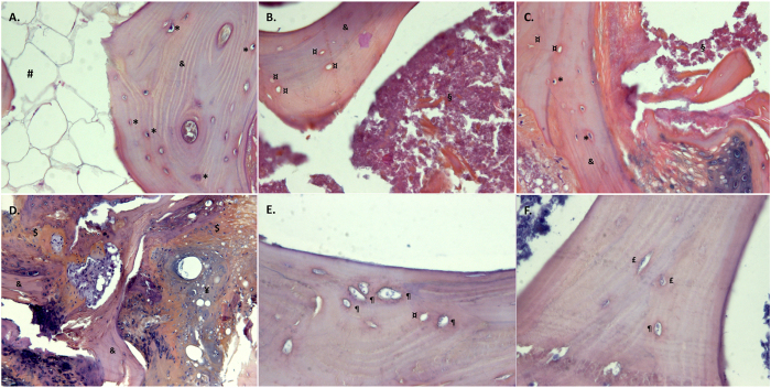
Figure 3.
Histological examination using hemtoxylin-eosin-saffron staining. (A) Control sclerotic-equivalent zone (x200); (B). Necrotic zone (ON group) (x200); (C). Sclerotic zone (ON group) (x200); (D). Sclerotic zone with coexisting normal trabeculae, woven and chondroid bone (ON group) (x200); (E). pyknotic apoptotic osteocytes (ON group) (x400); (F). coexistence of pyknotic osteocytes others presenting with nuclei showing spread and faded chromatin (x400). &Normal trabecula, $woven bone, ¥chondroid bone, #normal medullary space, §necrotic medullary space, *intact osteocyte, ¶apoptotic osteocyte, ¤empty osteocyte lacuna. ON: osteonecrosis.

