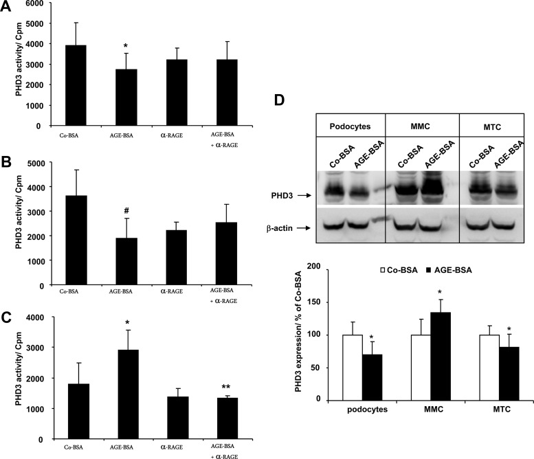
Figure 5.
A–D, PHD3 hydroxylase activity assay in renal cells treated with AGE-BSA. The cells were treated with 5 mg/mL Co-BSA or AGE-BSA for 24 hours. In an inhibitor study, the AGE receptor was blocked by addition of anti-RAGE antibody alone (α-RAGE) or together with AGE-BSA. An equal amount of protein lysate was subjected to the PHD3 hydroxylase activity assay using HIF-1α peptide as a substrate. A, PHD3 activity assay in differentiated podocytes. *, P < .05 vs Co-BSA (n = 8). B, PHD3 activity in MTCs. #, P < .01 vs Co-BSA (n = 8). C, PHD3 activity in MMCs. *, P < .05 vs Co-BSA; **, P < .001 vs AGE-BSA (n = 8). AGE-BSA treatment activates PHD3 prolyl-hydroxylase activity in MMCs and inhibits it in MTCs and podocytes relative to the cells treated with Co-BSA alone. D, Expression of PHD3 protein in differentiated podocytes, MMCs, and MTCs treated with Co-BSA or AGE-BSA analyzed by Western blot. A representative image of three independent experiments is shown. Upper panel, PHD3 Western blot; lower panel, β-actin protein expression as loading control. The graph presents a densitometry quantification of PHD3 protein expression in treated cells. The relative intensity of PHD3 protein is presented in percent relative to Co-BSA. *, P < .05 vs Co-BSA (n = 3).

