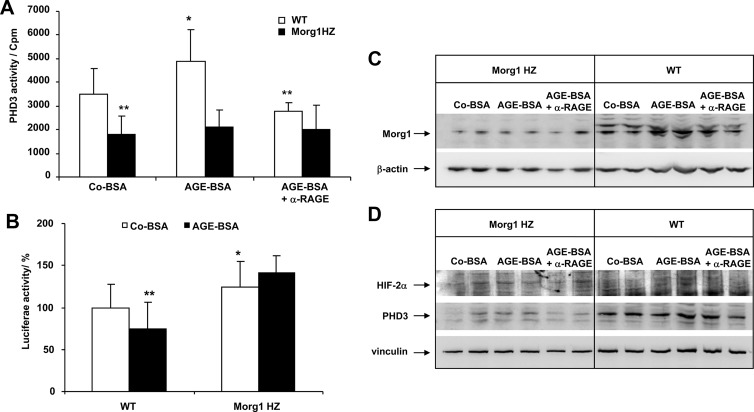
Figure 9.
A–D, Impact of AGE-BSA on PHD3 enzymatic activity and HIF-transcriptional activity in mesangial cells isolated from Morg1+/− HZ and WT (Morg1+/+) mice. A, PHD3 activity. AGE-BSA elevated the PHD3 enzymatic activity in mesangial cells isolated from WT compared with Co-BSA-stimulated cells. A significantly reduced PHD3 activity in Co-BSA as well as in AGE-BSA-treated mesangial cells obtained from Morg1+/− (Morg1 HZ) was measured relatively to WT mice. *, P < .05 vs Co-BSA WT; **, P < .005 vs Co-BSA WT (n = 9). Inhibitory studies demonstrated that PHD3 activity was regulated via the RAGE/AGE axis in WT cells. **, P < .005 vs AGE-BSA without α-RAGE (n = 9). In Morg1 HZ, AGE-BSA treatment failed to significantly induce PHD3 activity. B, Analysis of HIF-transcriptional activity by luciferase activity assay in WT and Morg1 HZ mesangial cells treated with Co-BSA and AGE-BSA (n = 18). HIF-luciferase reporter gene assays showed that AGE-BSA application to the WT mesangial cells significantly reduced HIF-DNA binding activity. In contrast, HIF-transcriptional activity was significantly increased in Morg1 HZ mesangial cells compared with WT cells. *, P < .05; **, P < .01 (n = 18). C, Protein lysates from Co-BSA, AGE-BSA, or AGE-BSA + α-RAGE antibody-treated mesangial cells obtained from Morg1 HZ or WT mice were subjected to Western blot analysis to monitor protein expression. Upper panel, Morg1 Western blot; lower panel, protein loading was controlled by detection of the β-actin protein expression. Representative Western blots are shown (n = 2 independent experiments). D, Protein expression of PHD3 and HIF-2α. The cells were treated as shown here and protein lysates were analyzed for protein expression. Upper panel, HIF-2α protein expression; middle panel, PHD3 protein Western blot; lower panel, detection of vinculin expression as a marker for protein loading. Clearly, less Morg1 and PHD3 protein expression was found in Morg1 HZ cells and no stimulation occurred with AGE-BSA. In contrast, AGE-BSA induced an increase in Morg1 and PHD 3 protein expression in WT cells that was reversed with the α-RAGE antibody. However, no major changes were seen in HIF-2α expression among the experimental conditions, probably due to the fact that cytoplasmatic lysates were used.

