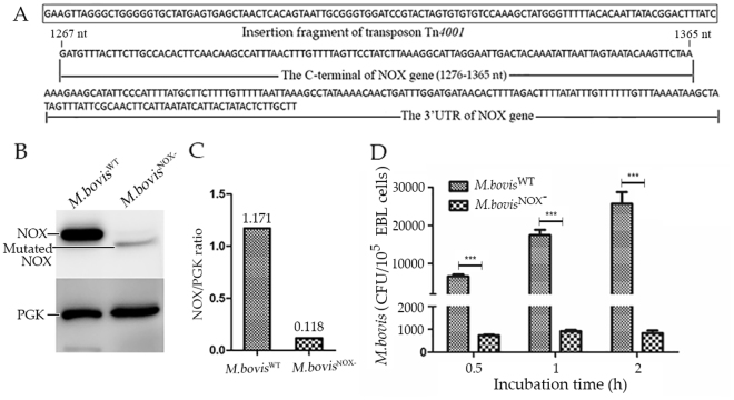
Figure 5.
Adhesion of M. bovis NOX− to EBL cells. (A) The result of sequencing Tn4001. The terminal sequence of Tn4001 was used for the amplification of sequence next to it. Then the sequence was used to map transposon insertion site and confirmed the NOX gene was inserted at 1276 nt (C-terminal of NOX gene). (B) The expression of NOX in M. bovis NOX−. The total proteins of M. bovis NOX− and M. bovis WT were incubated with antiserum against rPGK (1:500) or mAb (1:1000) to rNOX to detect the expression of NOX gene. The cropped blots are displayed here, the full-length blots are presented in supplementary information (Fig. S5). (C) Image J software was used to calculate the NOX/PGK ratio and then the expression of disrupted nox gene in M. bovis NOX− was compared with that intact nox in M. bovis WT. (D) The adhesion rates of M. bovis NOX− at three time points with MOI of 1:1000. ***p < 0.001 represents very significant difference.

