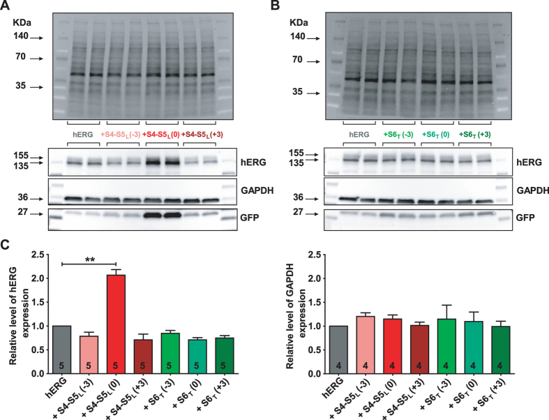
Figure 3.
S4-S5L (0) peptide increases hERG channel expression. (A,B) western blot analysis of hERG protein expression in COS-7 cell lysates in the absence or presence of S4-S5L peptides (A) or S6T peptides (B). Top: stain-free image of total proteins. Bottom: western blot images of hERG, GAPDH, and GFP proteins. The three blots, realized on the same membrane, are cropped. Full-length blots of each tested protein are reported in Supplemental Fig. 1. (C) Histogram of normalized mean intensity of hERG (left) and GAPDH (right) bands in the absence and in the presence of various S4-S5L or S6T peptides. Band intensities are first normalized to the intensity of the corresponding stain-free membrane lane, and ratios are then normalized to control hERG condition. **p < 0.01 versus control hERG, Mann-Whitney test realized on non-normalized ratios.

