Figure 2.
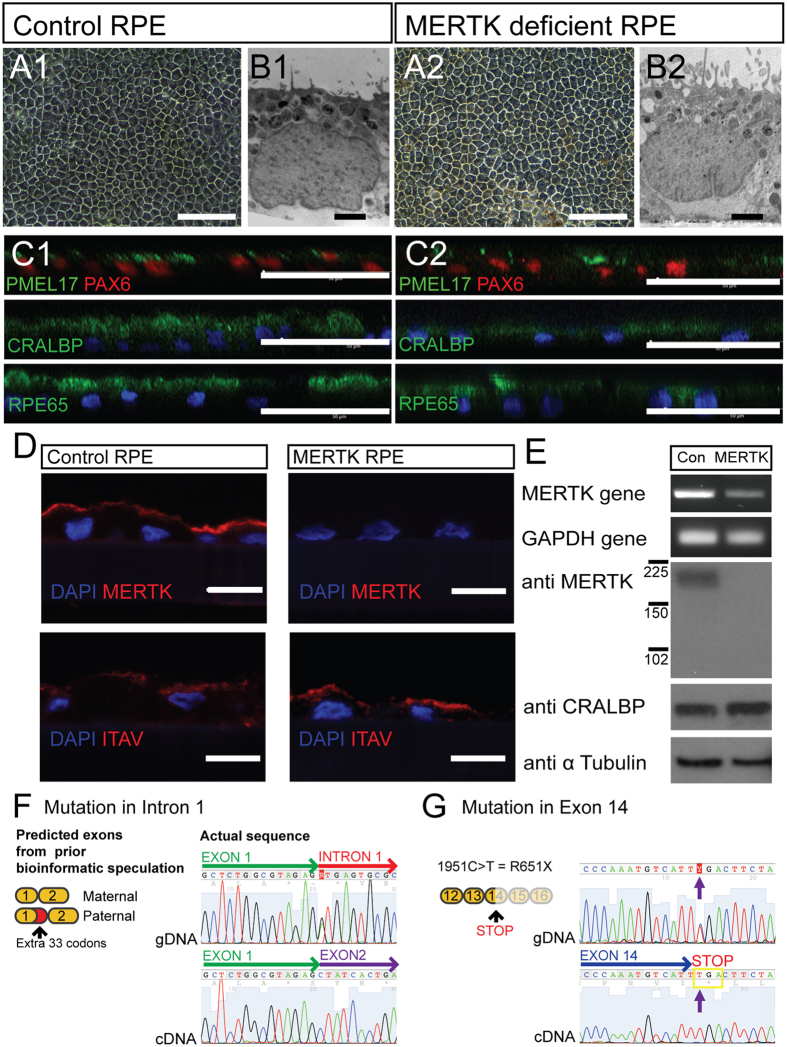
Characterisation of control and MERTK-RPE. (A1,2) Light micrograph of control and MERTK-RPE cells in culture (scale bar 50 μm). (B1,2) Electron micrograph of control and MERTK-RPE (scale bar 1 μm). (C1,2) Immunocytochemistry of iPSC derived RPE (scale bar 40 μm). (D) Immunohistochemistry of the control RPE and MERTK-RPE (scale bar = 10 μm). (E) RT PCR gel electrophoresis and Western blot of the MERTK gene and protein in the control RPE and MERTK-RPE. (F) gDNA sequence from MERTK fibroblasts of the exon 1/intron 1 junction of the MERTK gene compared to the corresponding MERTK-RPE derived cDNA sequence at the exon 1/exon 2 border. (G) gDNA sequence of MERTK fibroblasts compared to MERTK-RPE cDNA sequence in exon 14 of the MERTK gene.

