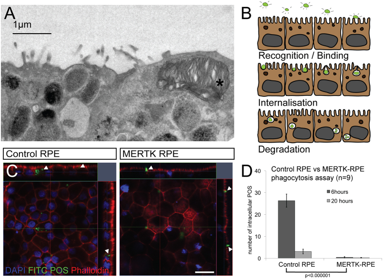
Figure 3.
MERTK-RPE are unable to phagocytose. (A) Electron micrograph of the apical surface of the control RPE 4 hours following the addition of isolated photoreceptor outersegments (POS) *. (B) Schematic of the process of POS phagocytosis by RPE using FITC labeled POS. (C) Orthogonal representations of confocal Z stacks of the control RPE and MERTK-RPE 6 hours following the addition of FITC labeled POS (scale bar = 20 μm), arrows highlight POS. (D) Graphical representation of phagocytosis between control RPE and MERTK-RPE at 6 and 20 hours (error bars represent the mean ±1SEM and p value derived from students t-test).

