Figure 2.
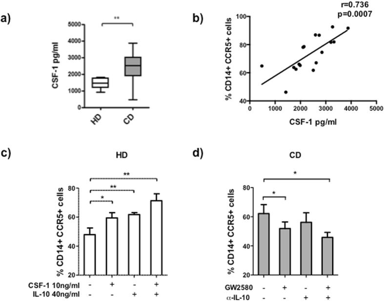
CSF-1 and IL-10 upregulated CCR5 expression on monocytes from HD and CD patients in remission. (a) The levels of CSF-1 after 24 h of WB culture of peripheral blood monocytes from HD (n = 14) and CD patients in remission (n = 17) were quantified by ELISA. (b) Correlation between CSF-1 levels and the percentage of CD14+CCR5+ peripheral blood monocytes from CD patients after 24 h of WB culture. (c) Peripheral blood monocytes from HD after 24 h of culture with medium, CSF-1 (10 ng/ml), IL-10 (40 ng/ml), or CSF-1+IL-10 were analyzed by flow cytometry. The results are expressed as a percentage of CD14+CCR5+ cells. (d) Peripheral blood monocytes from CD patients after 24 h of culture in the presence of medium, GW2580 (35 ng/ml), anti-IL-10 (3.5 ng/ml), or GW2580+anti-IL-10 were analyzed by flow cytometry. The results are expressed as a percentage of CD14+CCR5+ cells. Mann-Whitney U test was used for the comparison of CSF-1 concentrations between HD and CD patients. Spearman test was used to calculate the correlation between CSF-1 levels and CD14+CCR5+ cells. ANOVA was used for multiple comparisons of migration in HD (p < 0.0001) and CD (p = 0.05). Wilcoxon test was used for the comparisons between medium and each different condition in HD and CD. (*p < 0.05, **p < 0.01, ***p < 0.001).

