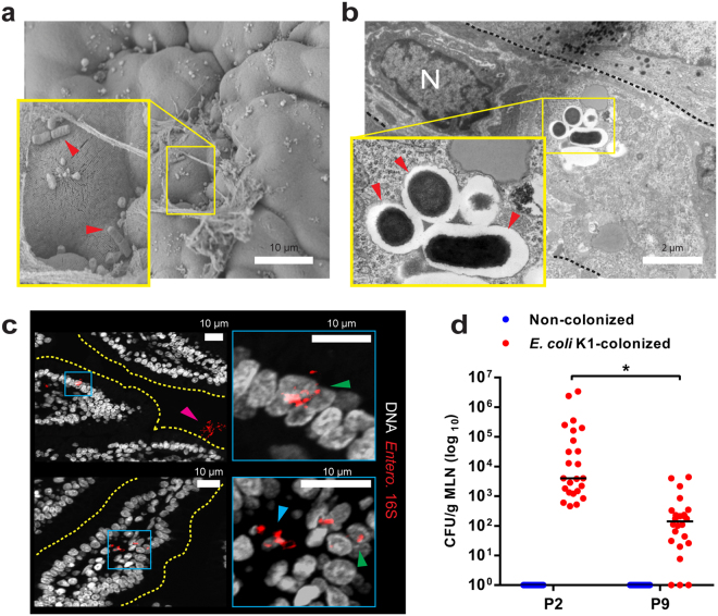
Figure 4.
E. coli K1 invasion of the MSI epithelium results in translocation of live bacteria to the intestinal mesenteric lymph nodes (a) SEM of MSI tissue from neonates 24 h after E. coli K1 colonization at P2; tissue-associated bacteria indicated ( ). (b) TEM of MSI enterocytes 24 h after E. coli K1 colonization at P2; intracellular bacteria within vesicles (
 ), enterocyte nucleus (N) and cell border (black dashed line) are indicated. (c) Confocal micrographs of MSI sections from neonatal rats 24 h after E. coli K1 colonization at P2; sections stained for Enterobacteriaceae 16S rRNA (red) and DNA (grey); right panel shows magnified images of regions defined by blue insets in left panels; luminal (
 ), intracellular (
 ) and translocated (
 ) bacteria are indicated. (d) Bacterial load within mesenteric lymphatic node tissue (MLN) harvested from P2 and P9 rats 24 h after initiation of colonization with E. coli A192PP, showing enumeration of E. coli K1 (phage K1E-susceptible). n = 23 animals for non-colonised and n = 24 animals for colonized pups; line is median, *P < 0.05 (Student’s t).

