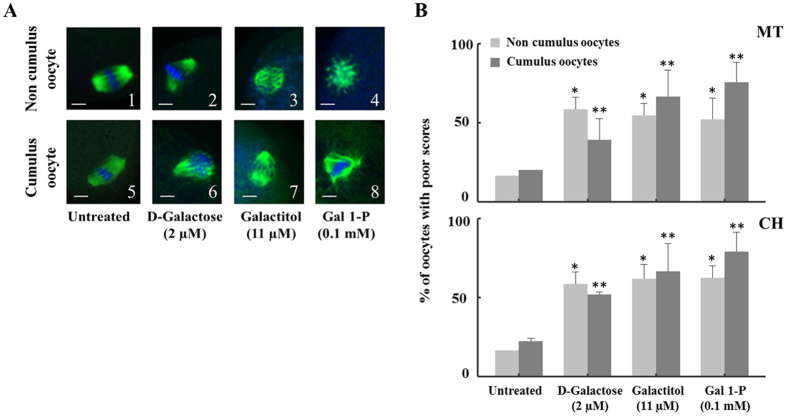
Figure 3.
The effect of galactose and its metabolites on Metaphase II mouse oocyte spindles and chromosomes. (A) Representative confocal images of non-cumulus and cumulus metaphase II mouse spindles stained with β-tubulin antibody to visualize the microtubules (MT) (green) and counterstained with DAPI to visualize chromosomes (CH) (blue). After 4 hours of incubation, various abnormal configurations of spindles were observed when oocytes were exposed to D-galactose (B,F), galactitol (C,G) or Gal 1-P (galactose 1-phosphate) (D,H) compared to normal spindle shapes in untreated group (A,E) (n = 30/group). Scale bars: 1 pixel, 3 mm. Images shown are from a typical triplicated experiment. (B) The percentage of oocytes with poor scores in MT structure (upper panel) and CH alignment (lower panel) (120 cumulus and 120 without cumulus) in untreated oocytes compared to oocytes treated with galactose, galactitol and Gal 1-P (galactose 1-phosphate). Poor scores were significantly increased in oocytes exposed to galactose and its metabolites compared with controls in both MT and CH, indicated by *for oocytes without cumulus cells (p = 0.032 for MT and p = 0.05 for CH) and **for oocytes with cumulus cells (p = 0.04 for MT and p = 0.029 for CH). The experiment was conducted in triplicate.

