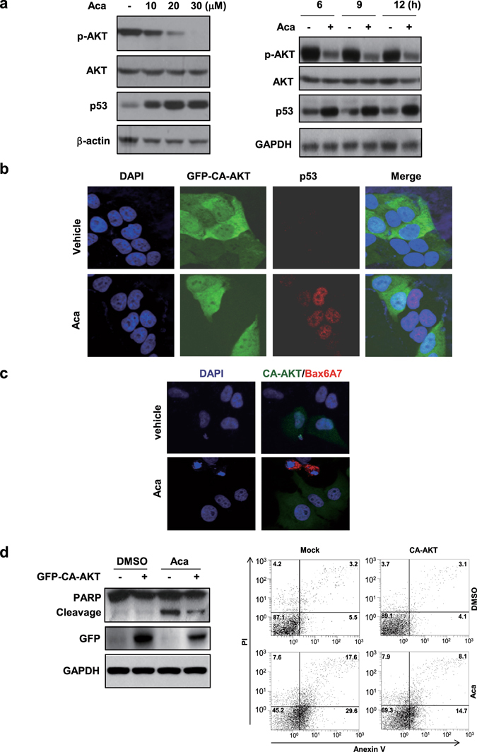
Figure 5.

AKT is a potent inhibitor of p53. (a) HepG2 cells were treated with different concentrations of acacetin for 6 h or with 15 μM acacetin for different time intervals. The inverse relationship between phosphorylated AKT and p53 was shown in the Western blotting. (b) HepG2 cells were transfected with constitutively active AKT (GFP-CA-AKT) and treated with 15 μM acacetin for 12 h. The cells were then immunostained with anti-p53 antibody and co-stained with DAPI. (c) HepG2 and its GFP-CA-AKT-transfected cells were treated with 15 μM acacetin for 12 h and then subjected to immuno-staining with conformation-specific Bax/6A7 antibody. The cells were co-stained with DAPI. (d) HepG2/CA-AKT and HepG/Mock stable cell lines were treated with 10 μM acacetin for 24 h. The cells were then subjected to immunoblotting (left) or Flow cytometry assays (right). All blots were cropped to remove irrelevant or empty lanes.
