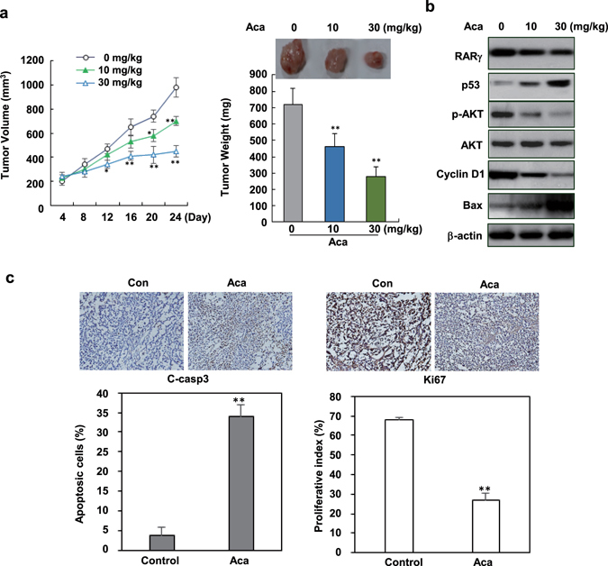
Figure 6.

In vivo anti-tumor activity of acacetin. (a) Mice were subcutaneously transplanted with HepG2/RARγ liver cancer cells. After 1 week, mice with palpable tumors were treated with 10 mg/kg (n = 6) and 30 mg/kg acacetin (n = 6) or vehicle (n = 6) by i.p. once every other day. Tumor volume was examined every 4 days after treatment. 3 weeks later of post-treatment, the mice were sacrificed and the tumors were collected for further assays. Representative tumors were shown and the effects of acacetin on tumor weights were evaluated. (b) The tumors were lysed and immunoblotted for assaying the expression of RARγ, p53, Bax, Cyclin D1, and the total and phosphorylated AKT proteins. (c) The effects of acacetin on the expression of cleaved caspase 3 (indicator of apoptosis) and Ki67 (proliferative index) were conducted by immunostaining. All the slides were co-stained with hematoxylin. The apoptotic and proliferative cell numbers were quantitated. All blots were cropped to remove irrelevant or empty lanes.
