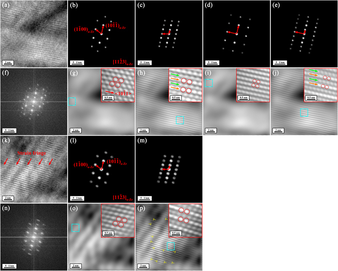
Figure 5.

Changes of atomic structure within the α-Zr matrix after irradiation at room temperature with focused and stationary electron beam at an incident energy of 30 keV in the FE-SEM viewed along the [113]α-Zr direction. (a) and (k) HRTEM images of two typical areas of the α-Zr matrix containing electron-beam-induced changes of atomic structure. (f) and (n) The FFT diffraction patterns corresponding to Fig. 5a and k, respectively. (b,c,d and e) The partially masked FFT diffraction patterns corresponding to Fig. 5f. (g,h,i and j) The noise-filtered IFFT images corresponding to Fig. 5a obtained by using the partially masked FFT diffraction patterns in Fig. 5b,c,d and e, respectively. The insets in Fig. 5g,h,i and j show the expanded morphologies of the areas outlined by blue squares in Fig. 5g,h,i and j, respectively. (l) and (m) The partially masked FFT diffraction patterns corresponding to Fig. 5n. (o) and (p) The noise-filtered IFFT images corresponding to Fig. 5k obtained by using the partially masked FFT diffraction patterns in Fig. 5l and m, respectively. The insets in Fig. 5o and p show the expanded morphologies of the areas outlined by blue squares in Fig. 5o and p, respectively. The yellow ‘T's in Fig. 5p indicate the electron-beam-induced dislocations.
