
Figure 4.
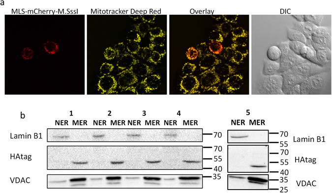
Mitochondrial localization of mitochondria-targeted DNA methyltransferases. (a) Confocal microscopy of HCT116 cells expressing MLS-mCherry-M.SssI. In order to stain the mitochondria, cells were incubated at 37 °C for 30 min. with 100 nM Mitotracker Deep Red. (b) Western blot of mitochondria-targeted M.CviPI, M.SssI or the catalytically inactive M.SssI ∆∆. Mitochondrial (MER) and nuclear (NER) protein extracts were isolated from C33A cells expressing mitochondria-targeted M.CviPI (lane 1) or M.SssI (lane 5) and HCT116 cells expressing mitochondria-targeted M.CviPI (lane 2), M.SssI (lane 3) or M.SssI ∆∆ (lane 4). A HAtag antibody was used to recognize the mitochondria-targeted constructs in the MER (49 kDa for M.CviPI, 52 kDa for M.SssI) or NER. Inside the mitochondria the mitochondrial-localization signal is cleaved off, reducing the size of the protein with 5 kDa. VDAC1/Porin (32 kDa) and Lamin B1 (68 kDa) were used as mitochondria and nuclear loading controls, respectively.