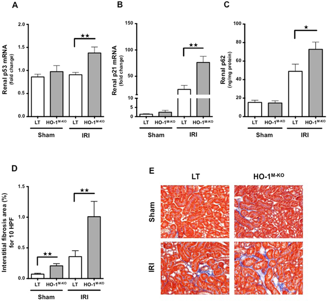
Figure 6.

Myeloid HO-1 promotes renal repair and attenuates interstitial fibrosis induced by renal IRI. (A) p53 mRNA, (B) p21 mRNA and (C) p62 renal expression in LT (white bars) and HO-1M-KO (grey bars) mice subjected to sham surgery, 4 hours (p53 mRNA analysis) or 24 hours (p21 mRNA and p62 analysis) of reperfusion after renal IRI. Results are expressed as the mean ± SEM, ★p < 0.05; ★★p < 0.01. In p53/p21 experiments, n = 6–8 for IRI groups and n = 5–6 for sham groups. In p62 experiment, n = 9–10 for IRI groups and n = 6 for sham groups. (D) Percentage of fibrosis area and (E) representative renal sections of Masson Trichrome staining from sham-operated and IRI after 7 days of reperfusion in LT (white bars) and HO-1M-KO (grey bars) mice. Quantification was done using ImageJ software. Results are expressed as the mean ± SEM, ★★p < 0.01. n = 3–4 for IRI groups and n = 5 for sham groups. Magnification, x200.
