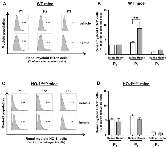
Figure 7.

Hemin specifically induces HO-1 expression in renal CD11b+ F4/80lo (P2) myeloid cells. WT (A,B) and HO-1M-KO mice (C,D) were treated with hemin (5 mg/kg) or saline. Twenty-four hours after intraperitoneal injection, kidneys were harvested and homogenized for flow cytometry analysis. The renal myeloid cell populations were characterized according to the expression of CD11b and F4/80 surface markers (i.e., CD11b− F4/80+ (P1), CD11b+ F4/80lo (P2), and CD11bhi F4/80-(P3)). (A,C) Representative histograms depicting the level of intracellular HO-1 expression in the myeloid subpopulations after hemin administration or saline injection. (B,D) Quantification of renal myeloid HO-1+ cells, presented as a proportion of the myeloid cells extracted from the kidney after hemin (grey bars) or saline (white bars). Results are expressed as the mean ± SEM, ★★p < 0.01. n = 7–8 per group in WT mice and n = 5 per group in HO-1M-KO mice.
