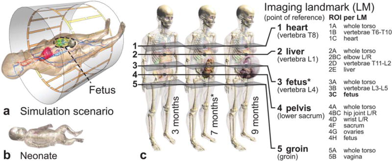
FIG. 1.

(a) Simulation scenario within the reference birdcage. (b) Neonate model included in the study. (c) Three anatomical pregnant women models at the third, seventh, and ninth month of gestation, with the evaluated imaging landmarks and corresponding ROIs per landmark. Many points of reference and ROIs are based on the locations of specific thoracic (T) and lumbar (L) vertebrae.
