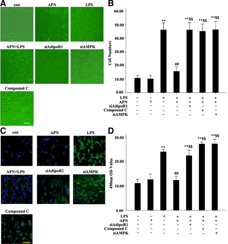
Fig. 7.

Effect of siAdipoR1, siAMPK, and AMPK inhibitor compound C on APN-reduced proliferation, migration and transformation of AFs. A, Scratch-wound assay results. B, Migration assay results. C, Immunofluorescent staining results of α-SM-actin. LPS, cells treated with LPS (10μg/ml) for 24 h; APN + LPS, cells pretreated with APN (10 μg/ml) for 2 h, then LPS (10 μg/ml) for 24 h; siAdipoR1 + APN + LPS, AFs treated with siAdipoR1, then with APN (10 μg/ml) for 2 h, then LPS (10 μg/ml) for 24 h; siAMPK + APN + LPS, cells treated with siAMPK (10 μm), then with APN (10 μg/ml) for 2 h, then LPS (10 μg/ml) for 24 h; compound C + APN + LPS, cells treated with compound C (20 μm) for 30 min, then with APN (10 μg/ml) for 2 h, then LPS (10 μg/ml) for 24 h. D, MTT assay results. **, P < 0.01 vs. control group (con); ##, P < 0.01 vs. LPS; $$, P < 0.01 vs. APN + LPS. Values are means ± sem.
