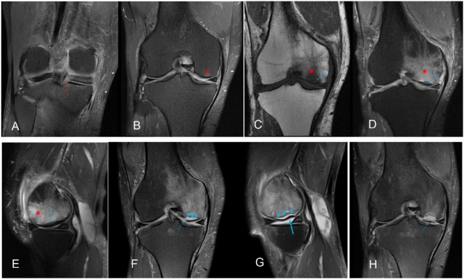
Figure 2.
A 56-year-old woman with pain on the medial interline for 3 months. (A) Coronal T2-weighted fat shows a radial tear of the posterior root of the medial meniscus (red arrow). (B) Coronal T2-weighted fat shows discrete subchondral edema of the condyle load-bearing area and medial tibial plateau, without detection of the subchondral fracture (asterisk). (C, D, E, F). Imaging performed 6 months later showing the classic findings of subchondral fracture, including edema (asterisk) and subchondral fracture (blue arrow). (F) and (G). After 12 months, signs of impaction/collapse (thick arrow) and subchondral cortical bone discontinuity (arrow) are evident; intra-articular fluid seeped into the fracture, which now appears as a line with high signal intensity on T2-weighted imaging (arrowhead). (H) After 18 months, the osteochondral fragment is fully detached.

