
Figure 1.
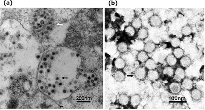
Ultrastructural characteristics of NDiV-SZ11706Z. (a) Ultrathin sections of NDiV in a cytoplasmic vacuole (black arrow) and a higher magnification of vesicles containing numerous virions (white arrow). (b) Electron micrograph of negatively stained NDiV virions.