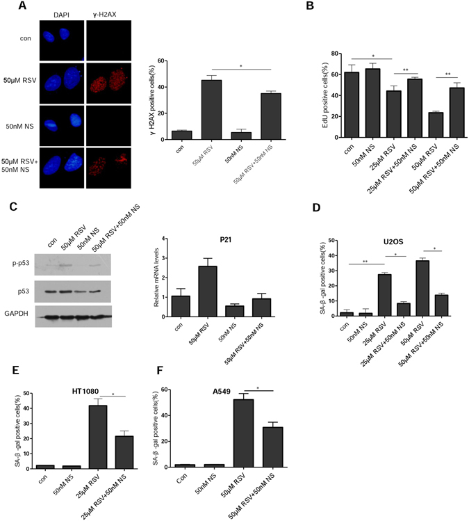
Figure 2.

Resveratrol-induced cellular senescence can be attenuated by nucleosides. (A) Immunofluorescence staining of γ-H2AX in U2OS cells. Cells growing on coverslips in 6-well plates were exposed to RSV (50 μM) alone or in combination with nucleosides (50 nM) for 24 h, and fixed for examination by immunofluorescence. (B) EdU proliferation assay was performed in U2OS cells 48 h after treatment with RSV alone or in combination with nucleosides, the percentages of EdU positive cells are shown as the mean ± S.D. from three independent experiments. (C) Left, U2OS cells were exposed to RSV with or without nucleosides for 48 h. Whole cell lysates were analyzed by immunoblotting with antibodies specific for p-p53 and p53, respectively. GAPDH was used as a loading control. Right, with the same treatment as in the left, but examined for expression of p21 mRNA levels by RT-PCR. (D) U2OS cells were incubated with RSV with or without addition of the indicated concentration of nucleosides (NS). On day 7, cells were examined for SA-β-gal activity. Mean of three independent experiments with SEM is shown. (E,F) The same as in (D) but in HT1080 and A549 cells, respectively. *P < 0.05, **P < 0.01.
