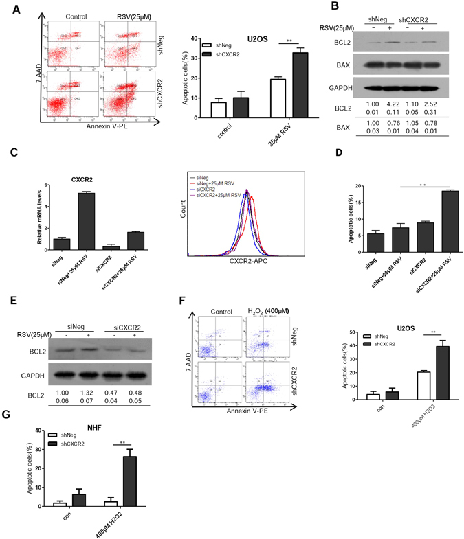
Figure 6.

CXCR2 protects cells from undergoing stress-induced apoptosis. (A) Apoptosis in shNeg and shCXCR2 U2OS cells was measured 3 days after treatment with RSV (25 μM) by flow cytometry. (B) Cells were treated as in (A) and whole-cell extracts were collected for Western blot analysis using BCL2 and BAX antibodies. (C) Downregulation of CXCR2 by siRNA as measured by RT-PCR and flow cytometry. U2OS cells were transfected with siRNA duplexes (200 nmol/L) specific to CXCR2 or scrabbled oligo in serum-free medium for 6 hours, then were incubated with complete medium for 24 h and then incubated with RSV for 3 days. (D) U2OS cells were treated the same as in (C) and apoptosis was measured by flow cytometry. (E) The U2OS cells were treated the same as in (C) and whole-cell extracts were collected for Western blot analysis using BCL2 antibodies. (F) Apoptosis in shNeg and shCXCR2 U2OS cells was measured by flow cytometry 2 days after treatment with H2O2 (400 μM). Results shown are representative of three independent experiments. (G) The shNeg and shCXCR2 NHF cells were treated the same as in (F) and apoptosis was measured by flow cytometry. The numbers shown below Western blot images are means (first row) and SE (second row) of band intensities relative to control. Signals on the immunoblots were analyzed by ImageJ, normalized with that of GAPDH.
