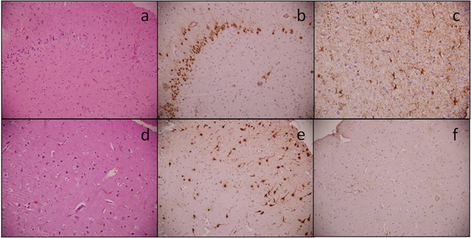
Figure 1.

Photomicrographs showing histopathology of the hippocampus and anterior temporal lobe. Representative pictures of the hippocampal specimen showing loss of neurons (a) HE, x200) highlighted by Neu N (b) IHC, x200), while GFAP shows reactive gliosis (c) IHC, x400). Anterior temporal lobe specimens show no loss of neurons (d) HE, x200 and (e) Neu N IHC, x200) or reactive gliosis (f) GFAP IHC, x200).
