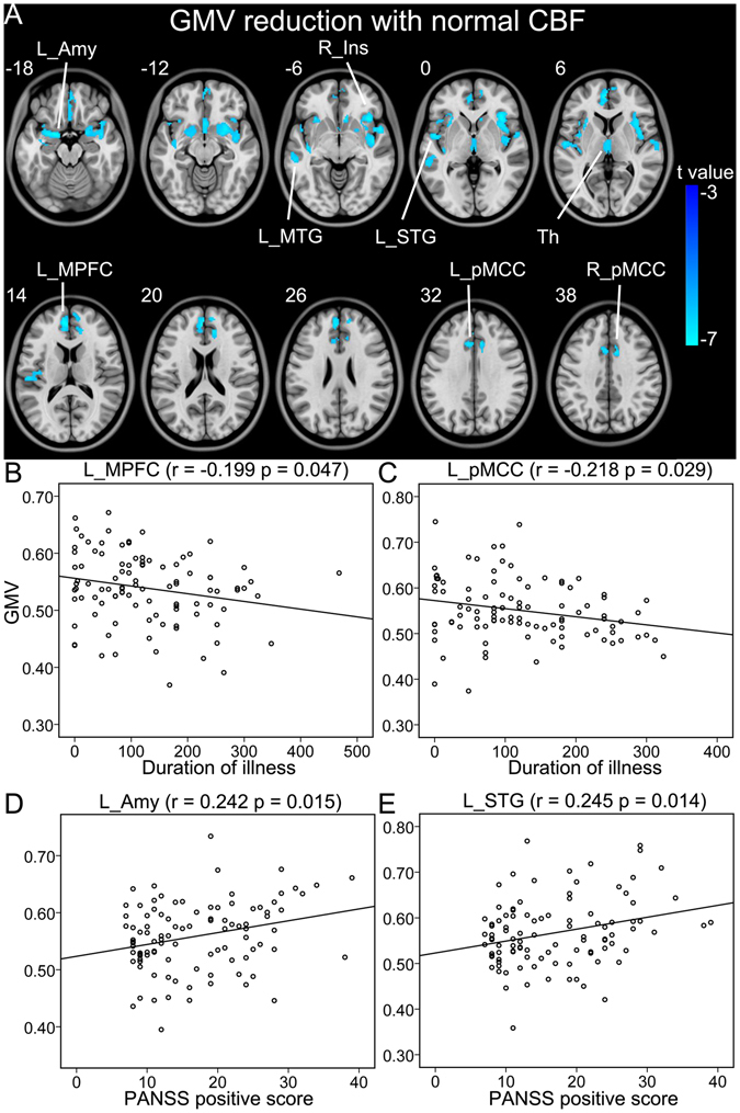
Figure 2.

Brain regions with reduced GMV and normal CBF in schizophrenia patients (A). (B–E) Shows correlations between GMV and CBF of these regions and clinical parameters in schizophrenia patients. Abbreviations: CBF, cerebral blood flow; GMV, grey matter volume; L, left; and R, right; Amy, amygdala; Ins, insular cortex; MPFC, medial prefrontal cortex; MTG, middle temporal gyrus; pMCC, posterior middle cingulate-cortex; STG, superior temporal gyrus. The r value represents the Spearman’s correlation coefficients.
