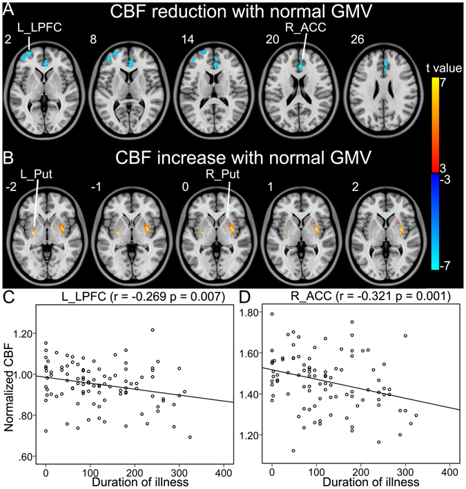
Figure 3.

Brain regions with normal GMV and abnormal CBF in schizophrenia patients. (A) Shows brain regions with normal GMV and reduced CBF in patients. (B) Shows brain regions with normal GMV and increased CBF in patients. (C,D) Show correlations between CBF of these regions and clinical parameters in schizophrenia patients. Abbreviations: CBF, cerebral blood flow; GMV, grey matter volume; L, left; and R, right; ACC, anterior cingulate-cortex; LPFC, lateral prefrontal cortex; Put, putamen. The r value represents the Spearman’s correlation coefficients.
