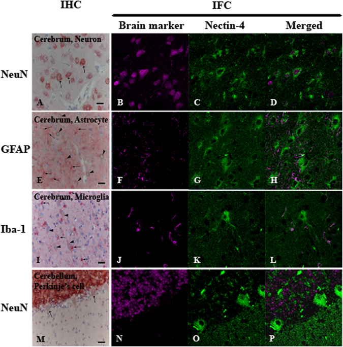
Figure 2.

Immunohistochemistry (IHC) and immunofluorescence assay (IFA) of nectin-4 expression in normal canine brain. Nectin-4 was stained in brown (IHC) and in green (IFA) while brain markers (NeuN, GFAP, Iba-1) was labeled in red (IHC) and in magenta (IFA). Merged panel of IFA revealed the co-localization of both antigens in particular cells. Neurons at cerebrum predominately co-expressed nectin-4 and NeuN (A–D, arrows). Astrocytes at cerebrum were stained with GFAP (E, arrows), not nectin-4 (E, arrow heads indicated neurons). Microglia at cerebrum were separately immunolabeled with Iba-1 (I, arrows) and nectin-4 (I, arrow heads indicated neurons). Purkinje’s cells at cerebellum showed weakly nectin-4 positive (M, arrows) without NeuN staining. (IHC, Bar = 20 μm; IFA, 4,000 fold magnification).
