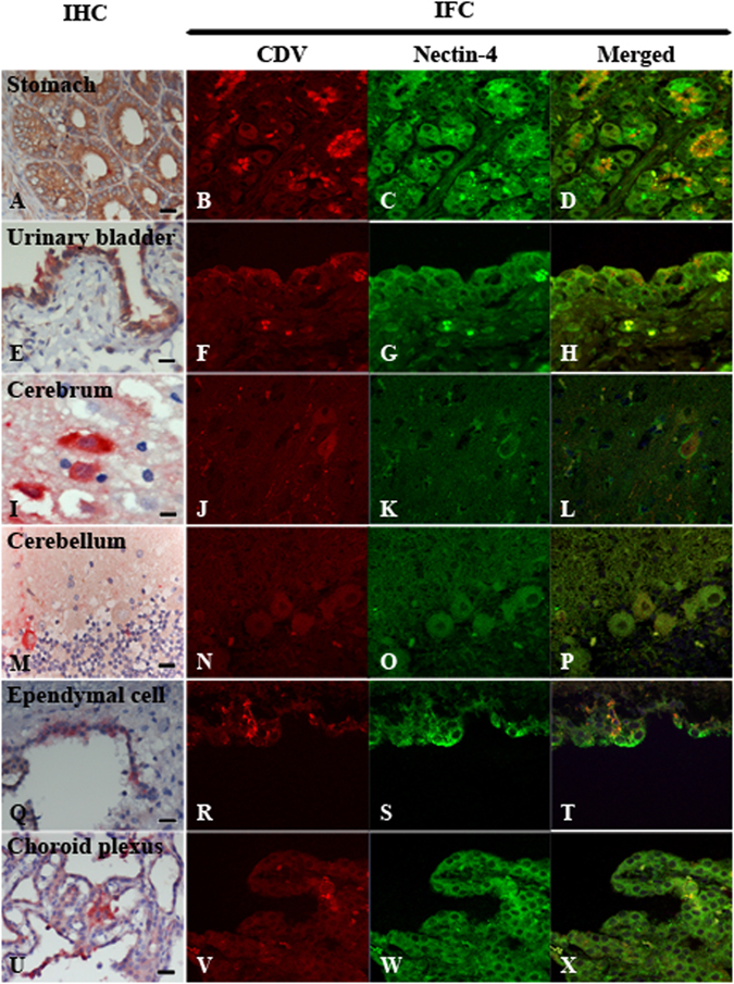
Figure 3.

Immunohistochemistry (IHC) and immunofluorescence assay (IFA) of nectin-4 expression in canine distemper virus (CDV)-infected tissues. Nectin-4 was stained in brown (IHC) and in green (IFA) while CDV antigen was showed in red (IHC, IFA). Merged panel of IFA revealed the co-localization of both antigens in particular cells. Non-nervous tissues including gastric glandular epithelial cells of stomach (A; data of case No. 2) and transitional epithelial cells of urinary bladder (E; data of case No. 6) were strongly positive for nectin-4 and CDV antigens. The visualization of co-expression was augmented when IFA was performed (B–D,F–H). In nervous tissues, neurons in cerebrum (I–L; data of case No. 10), Purkinje’s cells in cerebellum (M–P; data of case No. 13), ependymal cells (Q–T; data of case No. 11) and epithelial cells of choroid plexus (U–X; data of case No. 6) showed the co-expression of both antigens. (IHC, Bar = 20 μm; IFA, 4,000 fold magnification).
