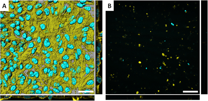
Figure 2.

Surface rendered confocal laser scanning microscopy (LSM) images of (A) the triple cell co-culture model cultured on a micro-porous membrane insert, and (B) a micro-porous membrane insert that originally had the co-culture system cultured upon it and subsequently having been treated with Trypsin-EDTA for ≥10 minutes at 37 °C, 5% CO2. Image (B) indicates that the Trypsin-EDTA was successful in detaching the majority of the co-culture from the micro-porous membrane insert in order to form a multi-cell suspension. Image A shows the morphology of the co-culture system when grown under normal culture conditions. Both images show staining for the F-actin cytoskeleton (Phalloidin-Rhodamine) and nuclear (DAPI) regions respectively. Scale bars represent 30 µm. In both (A and B), images show the XY plane in a 3D rendered format (as computed using the software IMARIS®, Switzerland), as well as planar views of XZ and YZ shown in 2D.
