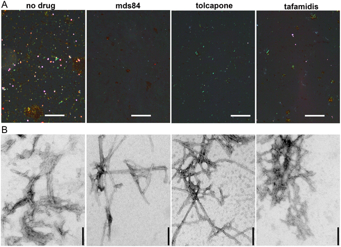
Figure 3.

Residual amyloid aggregates in the presence of excess of ligands. (A) Congo-red stained specimens viewed under intense cross polarized light in the absence of any ligands and in the presence of fourfold molar excess of mds84, tolcapone and tafamidis (Supplementary Fig. S6). Some fragments of amyloid are present with maximally inhibitory ligand concentrations (Fig. 1), although least with mds84. Scale bar, 100 µM. (B) Typical fibrillar structures detected by exhaustive analysis of negatively stained electron microscopy images of the same TTR-ligand preparations. Scale bar, 100 nm.
