


Figure 4.
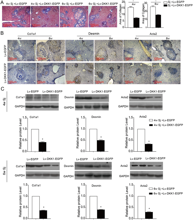
DKK1 transduction protects against schistosomiasis-induced liver fibrosis. Lv-DKK1-EGFP was injected via the tail vein to suppress Wnt signalling in the liver after S. japonicum infection for 4 or 6 weeks. (A) Masson’s trichrome staining was used to measure collagen fibre deposition, and the ratio of the collagen-stained area to the total area was also quantified. (B,C) Fibrotic markers (Col1a1, desmin, and α-SMA/Acta2) in the liver were assessed by immunohistochemical staining and western blotting. Sj, S. japonicum. Scale bar, 100 μm. Data represent the mean ± SEM; n = 5 (*P < 0.05).