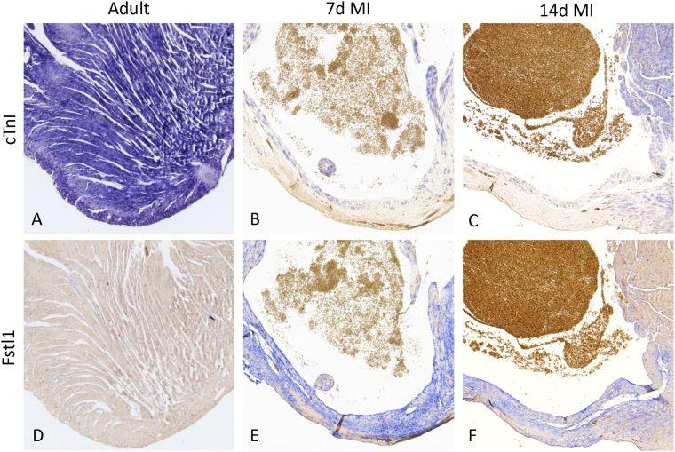
Figure 2.
Fstl1 expression in the mouse heart after myocardial infarction (MI). In situ hybridization on sections was performed to visualize the expression of cTnI and Fstl1 in control ventricle (A and D) and after induced myocardial infarction (B,E,C and F). The infarcted area is identified by the absence of cTnI expression (B and C). Fstl1 expression is low in control heart (D) and highly expressed in the infarcted area at 7 days (E) and 14 days (F) after myocardial infarction (7d MI and 14d MI).

