Figure 1.
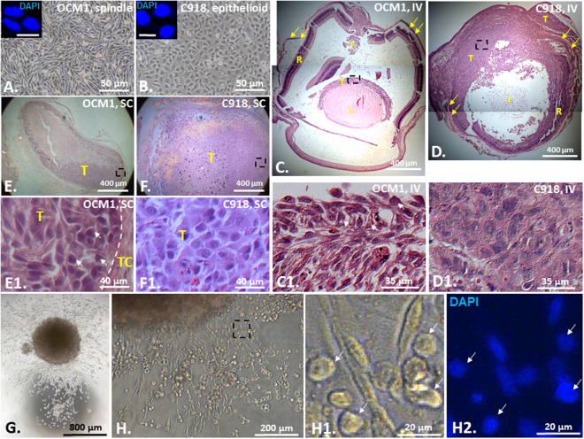
Epithelioid C918-derived tumors are more malignant than spindle OCM1-derived tumors. Confluent monolayer cultures of (A) the spindle UM cell line OCM1 and (B) the epithelioid UM cell line C918. Scale bar = 10 μm. Representative H&E stained images of both (C) OCM1- and (D) C918-derived ocular tumors generated in 13 days after IV injection, and both (E) OCM1- and (F) C918-derived cutaneous tumors generated in 13 days after SC injection. (G) A single OCM1 cell-formed sphere touched down and cells migrated out of the sphere. (H) OCM1 sphere-derived polymorphic cell types (inserts) including those with rounded epithelioid/amoeboid shape (arrows). IV = Intravitreal injection; SC = subcutaneous injection; L = Lens; R = Retina; T = Tumor; TC = Tumor Capsule. Yellow arrows indicate the sclera. White arrows indicate epithelioid cells in the OCM1-derived tumors and from an OCM1 sphere. Black dashed rectangles indicate the bottom-side inserts. Mean (M) ± standard deviation (SD).

