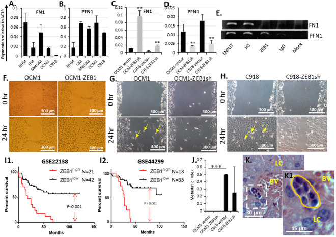
Figure 8.
ZEB1 facilitates UM cell migration. Transcriptional analyses showed that (A) the major component of extracellular matrix (ECM) fibronectin (FN1) and (B) the locomotion protein profilin (PFN1) were under- or over-expressed in UM, MetUM, and UM cell lines, respectively. Knockdown of ZEB1 increased (C) FN1 but decreased (D) PFN1 expression accordingly. (E) ChIP assays showed that ZEB1 bound to both FN1 and PFN1 promoters. Detected by scratch assays, (F) overexpression of ZEB1 in OCM1 increased cell migration while knockdown of ZEB1 in (G) OCM1 and in (H) C918 cells reduced their migration to cross the scratched gap. Yellow arrows indicate that semi-suspended cells detached from the monolayer culture are of amoeboid cell shape. (I) Kaplan-Meier survival curves of two large cohorts of total 63 and 53 primary UMs, respectively (microarray data sets GES22138 and GES44299, downloaded from NCBI database, Supplemental Tables 4 and 6), were generated and compared between ZEB1 high group (n = 21 or 18) and ZEB1 low group (n = 42 or 35). (J) Equal number of OCM1- and C918-vector controls and ZEB1sh knockdown cells (2 × 10⁵) were intravitreously injected into the nude mice and their 25-day grafted ocular tumors were examined for potential metastasis evaluation. (K) A representative image where potential metastatic cells are seen to be close to a blood vessel (BL) and surrounded by the normal liver cells (LC) 25 days after intravitreal injection of C918-vector control cells. Mean (M) ± standard deviation (SD). Student’s t-tests were performed. ‘**’ Indicates p ≤ 0.01. *** Indicates p ≤ 0.001. Yellow arrows indicate amoeboid-like cell morphology.

