
Figure 3.
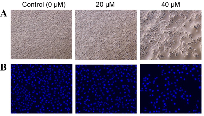
Morphological changes in HK-2 cells and their nuclei in response to POA treatment. (A) Phase contrast images of HK-2 cells following treatment with 0, 20 or 40 µM POA for 24 h (original magnification, ×40). (B) Fluorescence photomicrograph of HK-2 cells stained with Hoechst 33258 following treatment with 0, 20 or 40 µM POA for 24 h (original magnification, ×40). POA, oxalicumone A.