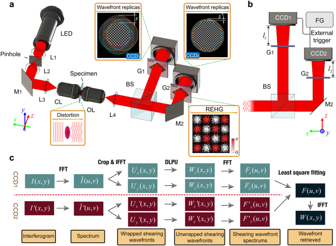
Figure 1.
Principle of WSEIM and our phase retrieval algorithm. (a) Optical layout of WSEIM: L1, L2, L3 and L4, lenses; M1 and M2, mirrors; CL, condenser lens; OL, objective lens; BS, beam splitter; G1 and G2, REHGs. The light emitted from a LED chip is collimated by a collimator first and filtered by a pinhole to become a plane wave probe when it reaches the specimen. The variation of refractive index distribution in the specimen results in the wavefront distortion of the probe. Then the probe is divided into two beams and each of them is diffracted into four replicas by a REHG. Two quadriwave lateral shearing interferograms with different lateral shear s and s′ are recorded by CCD1 and CCD2 simultaneously. (b) Schematics of the wideband-sensitivity-enhanced phase imaging system: FG, function generator. The difference in the lateral shears is controlled by altering the distances l 1 and l 2 from the REHGs to the CCD image plane. An external trigger signal provided by a function generator is employed to synchronize the image acquisition of two CCDs. (c) Schematic diagram of the phase retrieval algorithm for WSEIM. The first three steps are similar with the phase retrieval in the DHM, but the wavefronts obtained with these steps are just the shearing wavefronts. The differential leveling phase unwrapping (DLPU) algorithm is proposed to unwrap the shearing wavefronts. And the wavefront under test will finally be retrieved from its Fourier spectrum, which can be obtained from the four spectrums of shearing wavefronts using least-square fitting method.

