Figure 6.
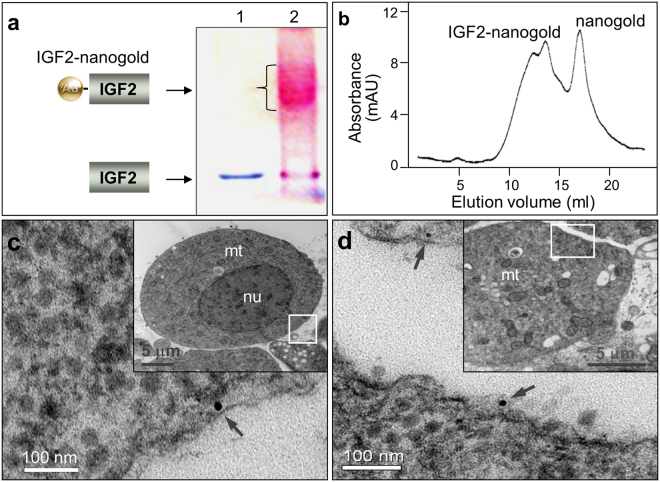
Location of IGF2 on the cell membrane. IGF2 was conjugated with nanogold and incubated with medaka blastomeres. Under electron microscopy, the ligand was detected on the cell surface or beneath the membrane. (a) 10% SDS-PAGE analysis of non-reduced and unboiled IGF2 (lane 1) and IGF2-Nanogold conjugate (lane 2). IGF2 was incubated with 1.4 nm nanogold, and the production was analyzed with SDS-PAGE. The protein band migrated at a higher apparent molecular weight after incubation with Nanogold (lane 2), demonstrating covalent attachment of 1.4 nm nanogold to IGF2. (b) Purification of IGF2-Nanogold conjugate with gel filtration. (c and d) Location of IGF2-nanogold under TEM, showing IGF2-nanogold (arrow) binding to cell. Insets depict the nucleus (nu), mitochondrial (mt) and location of enlarged areas of cells (boxes).

