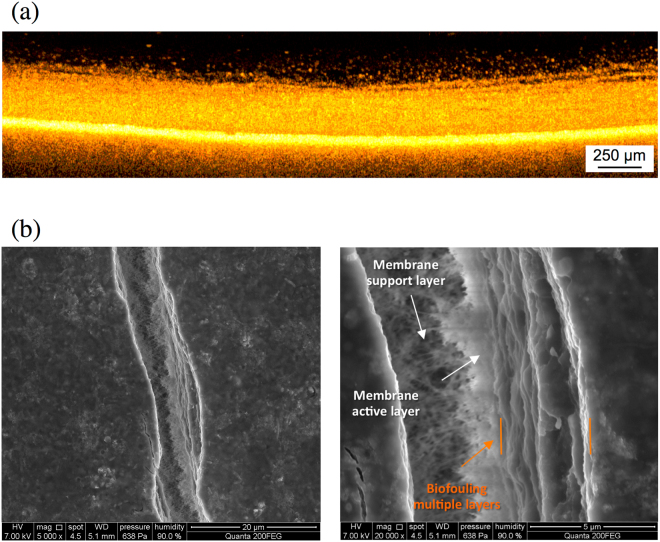
Figure 8.
(a) OCT cross-sectional biofouling analysis after 42 d. It is possible to observe the presence of biofouling multilayers above the membrane. (b) ESEM tilted surface image of the fouling layer after 42 d. The fracture on the surface was produced by gradually decreasing the humidity in the chamber.

