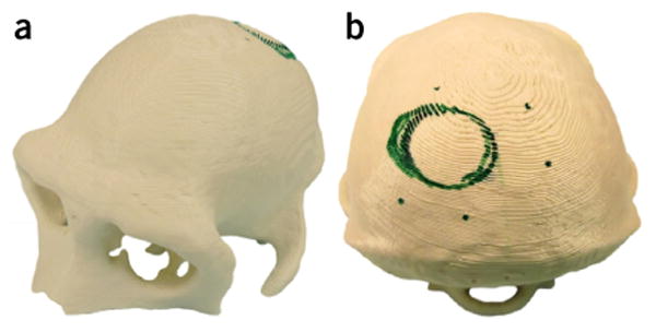
FIGURE 2.

Example 3D-printed model, made from MRI images of a macaque's skull and used for realistic surgical planning. (a) Oblique anterior view and (b) posterior view of the model. Green marker shows the planned location of the recording chamber (circle) and screws (dots).
