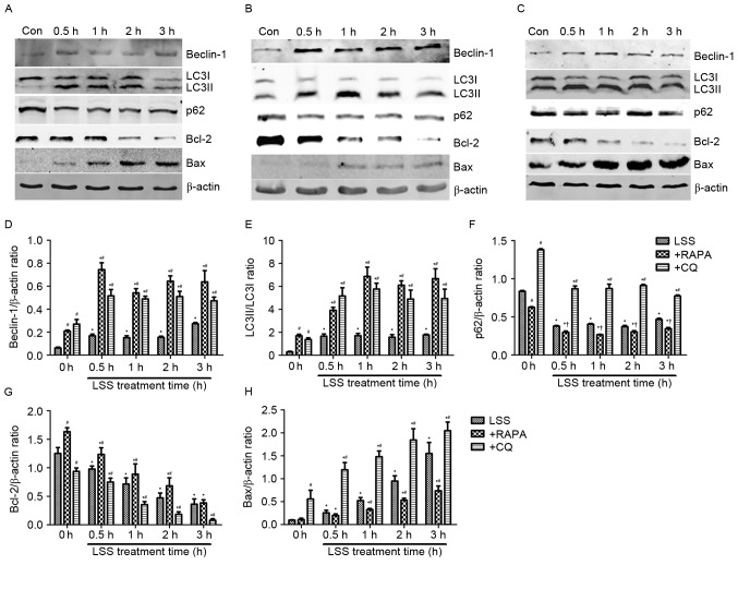
Figure 3.
LSS induces autophagy and apoptosis in HUVECs. The expression levels of the Beclin-1, LC3I, LC3II, p62, Bcl-2 and Bax proteins were determined by western blotting, following treatment with (A) LSS, (B) LSS+RAPA and (C) LSS+CQ. (D) Beclin-1 intensity, (E) LC3II/LC3I intensity, (F) p62 intensity, (G) Bcl-2 intensity and (H) Bax intensity were expressed as the fold change between LSS+RAPA, LSS+CQ and LSS. The bar graphs represent the mean ± standard error (n=3). *P<0.01 vs. the control. †P<0.05 and #P<0.01 vs. the cells pretreated with the various modulators at the same point. Con, control; LSS, low shear stress; HUVEC, human umbilical vein endothelial cells; RAPA, rapamycin; CQ, chloroquine; LC3, MAP1 light chain 3-like protein; Bcl-2, apoptosis regulator Bcl-2; Bax, apoptosis regulator BAX.

