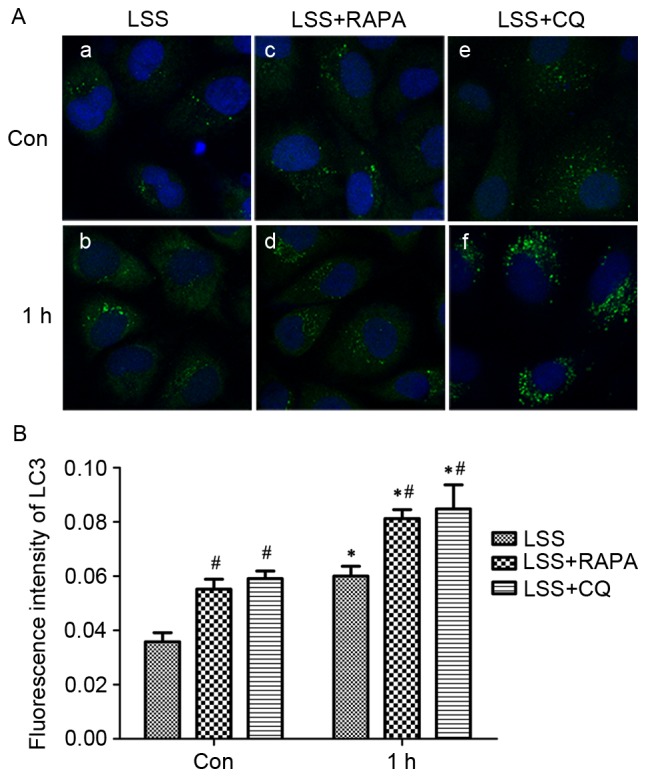
Figure 5.

LSS induced autophagosome formation in HUVECs. (A) Cells were maintained under static conditions as controls or subjected to LSS (1.5 dyn/cm2) for (a) 0 h or (b) 1 h. The cells were pretreated with RAPA (5 nM) for 24 h and subjected to LSS (1.5 dyn/cm2) for (c) 0 h and (d) 1 h. The cells were pretreated with CQ (20 µM) for 24 h and subjected to LSS (1.5 dyn/cm2) for (e) 0 h and (f) 1 h. The LC3 puncta (autophagosomes) in the cells were visualized by fluorescence microscopy after immunofluorescence staining with an LC3 antibody, followed by a fluorescein isothiocyanate-labeled secondary antibody (green, original magnification ×400). The nucleus was stained with DAPI (blue, original magnification ×400). (B) Quantification of fluorescence intensity of LC3. Bar graphs represent average number of typical LC3 puncta/cell. Data are mean ± standard error from minimum 30 cells for each experiment (n=3). *P<0.01 vs. the control. #P<0.01 vs. the cells pretreated with the various modulators at the same point. LSS, low shear stress; HUVEC, human umbilical vein endothelial cells; Bax, apoptosis regulator BAX; LC3, MAP1 light chain 3-like protein; RAPA, rapamycin; CQ, chloroquine.
