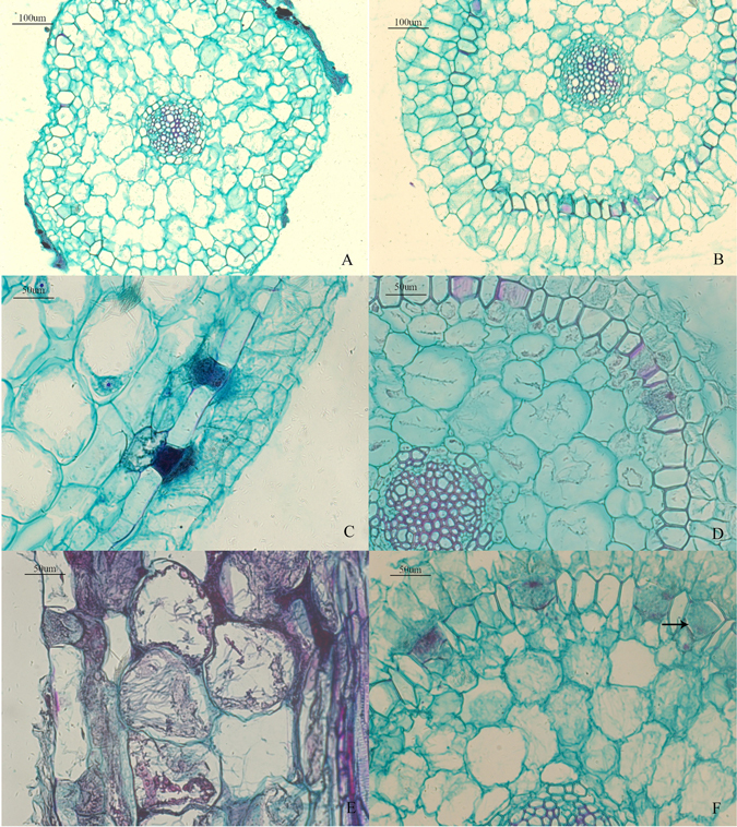
Figure 3.

Light micrographs of interactions between the roots of D. nobile and MF23 (A–F). (A) Transverse section of the control (×10). (B) Transverse section of the root after 1 week of symbiotic cultivation (×10). (C) Vertical section of the root after 3 weeks of symbiotic cultivation (×20). Distribution of a large number of hyphae outside the passage cells of the exodermis. (D) Transverse section of the root after 6 weeks of symbiotic cultivation (×20). The hyphae penetrated the cortex cell walls and colonized neighbouring cells through passage cells into the cortical region. (E) Vertical sections of the root after 6 weeks of symbiotic cultivation (×20). Hyphae passed through the cell wall to progress towards the other cortical cells. (F) Transverse section of the root after 9 weeks of symbiotic cultivation (×20). The peloton was formed in the passage cells.
