Figure 2.

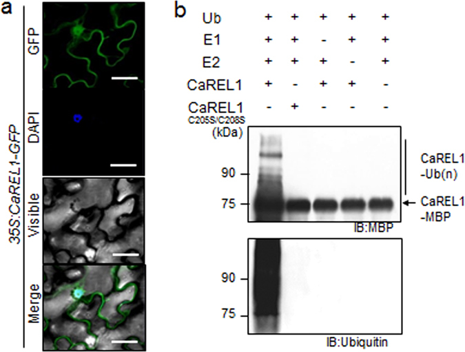
CaREL1 is localized at nucleus and has E3 ligase activity. (a) Subcellular localization of the 35S:CaREL1-GFP fusion protein in Nicotiana benthamiana epidermal cells. The 35S:CaREL1-GFP construct was expressed via agroinfiltration of N. benthamiana leaves and observed using a confocal laser-scanning microscope. DAPI staining was used as a marker for the nucleus. The scale bar represents 20 μm. (b) Auto-ubiquitination of CaREL1. In the presence of ubiquitin, E1 (UbE1), and E2 (UBHPC H5b), maltose-binding protein (MBP)–CaREL1 fusion proteins displayed E3 ubiquitin ligase activity. Detection of MBP–CaREL1 auto-ubiquitination. MBP–CaREL1C205S/C208S was loaded as negative control. MBP–CaREL1 fusion proteins were detected using MBP and ubiquitin antibodies; shifted bands indicate the attachment of ubiquitin molecules.
